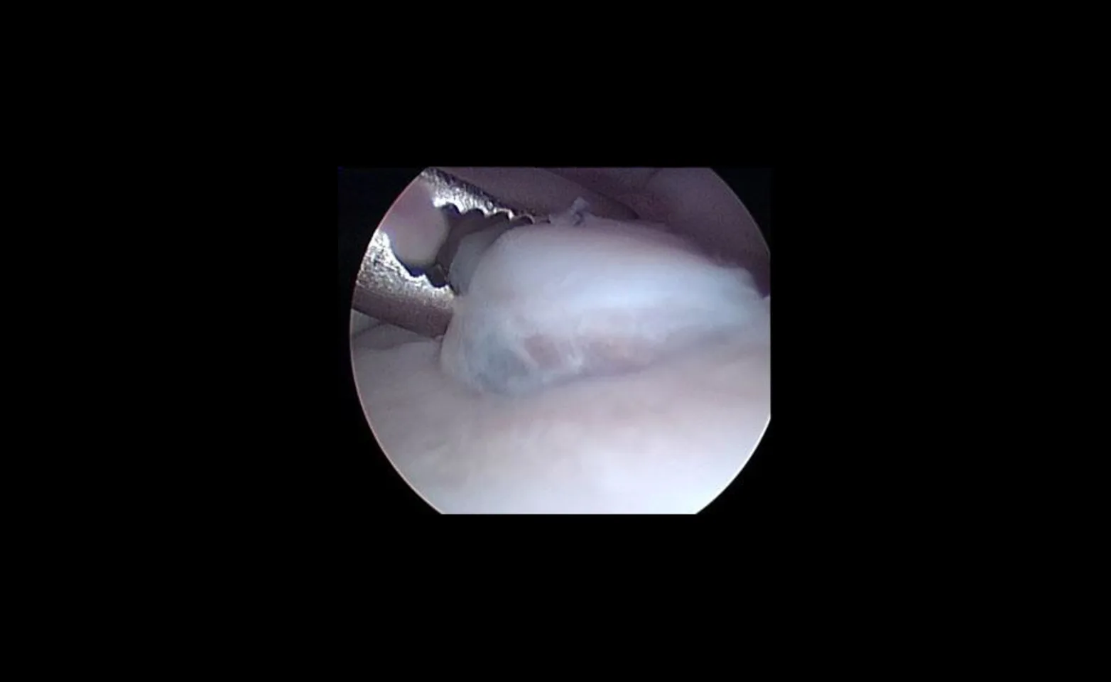
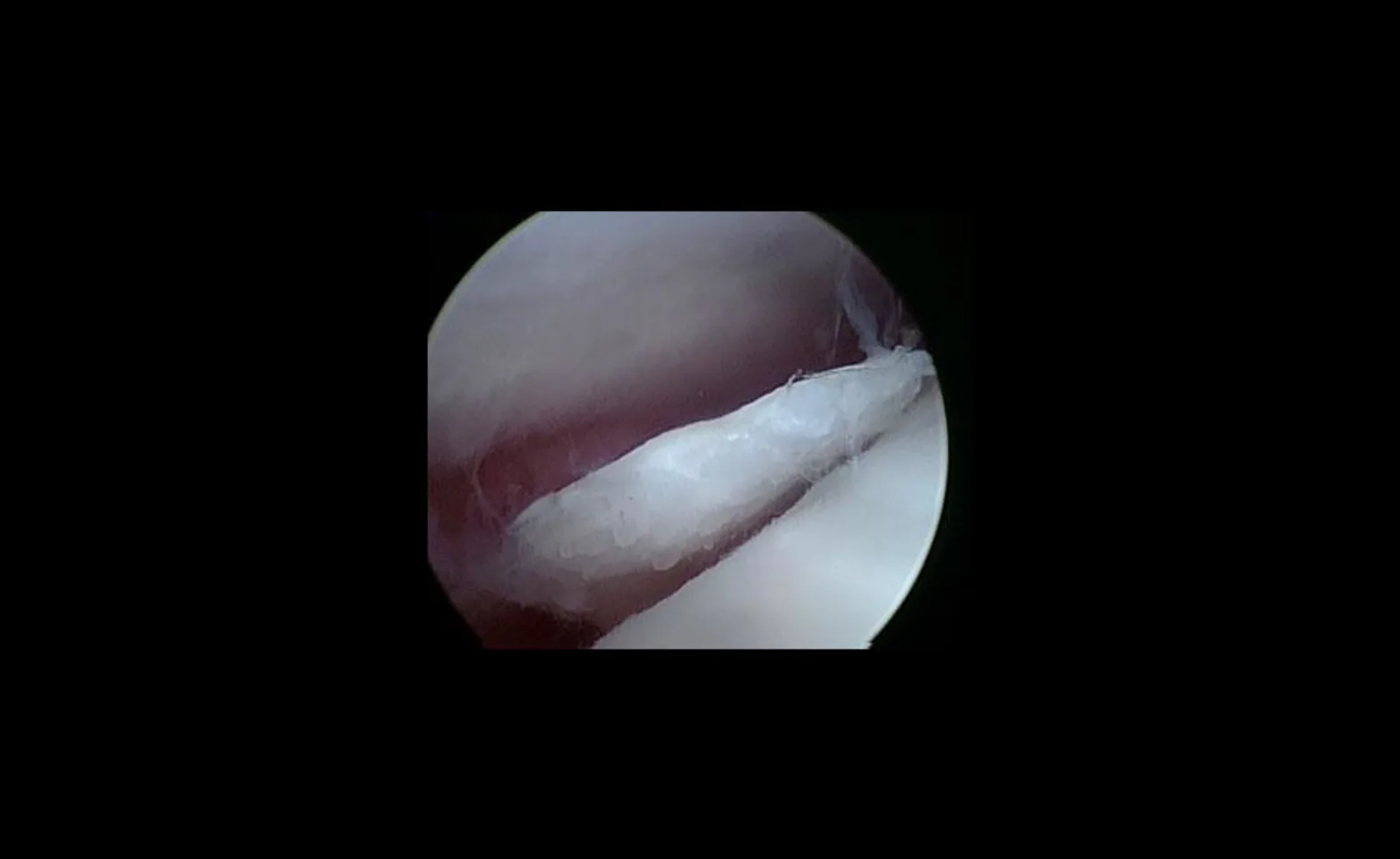
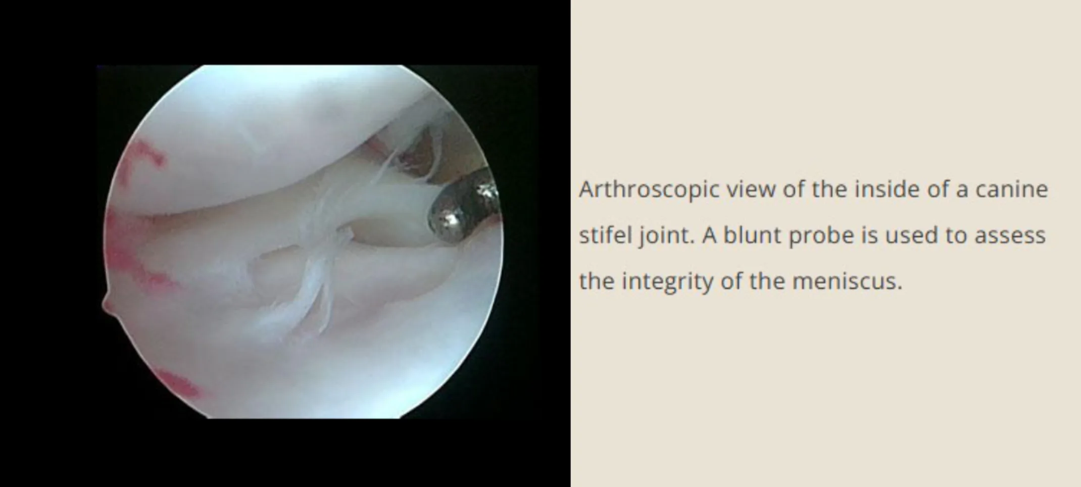
Verication and debridement of torn cranial cruciate ligament, identification, and treatment of meniscal injuries, stifel OCD, caudal cruciate ligament injuries.
Dallas Veterinary Surgical Center
Arthroscopy is for both the diagnosis and treatment of a variety joint diseases. Arthro- is derived from the Greek word for joint while –scope (“skopein”) is a Greek word meaning an instrument for viewing.
There are multiple advantages of arthroscopy over open arthrotomy with a traditional incision. Arthroscopy is minimally invasive and allows better visualization of the intraarticular structures and pathology. Patients generally have a quicker recovery and are less painful compared to arthrotomy patients. Additionally, arthroscopy allows for both the diagnosis and treatment of many conditions. From a medical perspective, arthroscopy allows for a more detailed evaluation of the joint due to the magnification that the equipment provides. Surgeons can accurately assess the degree and extent of articular cartilage damage. Another advantage is the ability to perform multiple joint procedures under one anesthesia.
For arthroscopy two, sometimes three, small 0.5-1.5cm incisions are made in the skin through which arthroscopic instrumentation is inserted. The joint is illuminated by light provided through a fiberoptic cable. A small camera is inserted into the joint that relays the images to a viewing screen which allows magnification and exceptional visualization of joint structures. Video and/or still picture images are saved for future reference and to allow for consultation with the owners in the postoperative period. Small additional portals are made to allow introduction of a motorized shaver and small hand instruments to allow for probing and smoothing of bone and cartilage surfaces, and for removal of loose or displaced bone chips or fragments.
After the inside of the joint has been evaluated and conditions treated, the instruments are removed and the incisions are closed with 1-2 skin sutures. During the surgery patients are kept under general anesthesia and immediately following the procedure a local anesthetic is injected into the joint to reduce discomfort. Postoperatively, patients may be kept in a soft padded bandage overnight depending on the joint evaluated. The small skin incisions heal within 7-10 days.

Elbow
Elbow dysplasia, fragmented coronoid process (FCP), humeral osteochondrosis dissecans (OCD), intraarticular fracture verification, microfracture technique for cartilage regeneration.
Arthroscopic view of a grasping instrument removing a large fragmented coronoid process within the elbow joint of a dog.

Shoulder
OCD of humeral head, rotator cu/medial compartment injury, damage to caudal glenoid cavity, biceps tendonitis, microfracture technique.
Arthroscopic view of a large cartilage ap in the shoulder of a dog, called caudal humeral osteochondritis dessicans.


