Dallas Veterinary Surgical Center

Visualizing lesions affecting the spinal cord has always been challenging, since many are not apparent on routine radiographs. We are frequently presented with patients who have complaints of ataxia, uncoordinated walking, weakness, paresis, or spinal pain. Choosing which imaging tests to perform can be confusing. Each form of imaging has its place and its own unique benefits and pitfalls.
Plain radiographs of the spine are useful in evaluating the vertebral column for the position of bones, continuity of the spinal column and spinal canal, bone density, changes in the articular facet joints, and the integrity of vertebral body end plates and disc spaces. Good positioning and radiographic technique are critical for evaluating the spine. Many lesions may be missed completely, and many anatomically normal things can be misinterpreted as pathology if good positioning and technique are not applied. This is particularly true if one is evaluating the width of the disc spaces. Sedation or general anesthesia may be required.
Lesions that may be readily diagnosed with plain radiographs of the spine include spinal fractures, luxations, and vertebral anomalies (such as hemivertebrae or spina bifida), discospondylitis, and some vertebral body tumors.
Because the spinal cord and discs cannot be visualized, plain radiographs are inadequate in the diagnosis of intervertebral disc extrusion, spinal cord or nerve root tumors, or congenital spinal cord defects (such as syringomyelia).

Myelography involves the injection of iodinated contrast into the subarachnoid space. Radiographs performed following this injection allow visualization of the outline of the spinal cord. Compressive lesions can be readily seen using this technique. Disc extrusion or extradural spinal cord tumors are commonly diagnosed with a myelogram. Vertebral instability, such as that seen in dogs with Wobbler’s syndrome, is most readily appreciated on myelographic views taken with the neck in several positions: neutral, flexion, or lateral distraction.
A myelogram does not allow us to evaluate the cauda equina for nerve root compression, such as is seen with lumbosacral stenosis, since the canine spinal cord ends around L5. Intramedullary spinal cord lesions, such as neoplasia or congenital syringomyelia, may be difficult to assess because the parenchyma of the spinal cord is not visualized.
Because it involves the injection of contrast media into the space around the spinal cord, a myelogram is an invasive procedure, and there is some associated risk. General anesthesia is required. Possible complications of myelography include transient post-myelogram seizures and worsening of the patient’s neurologic status.

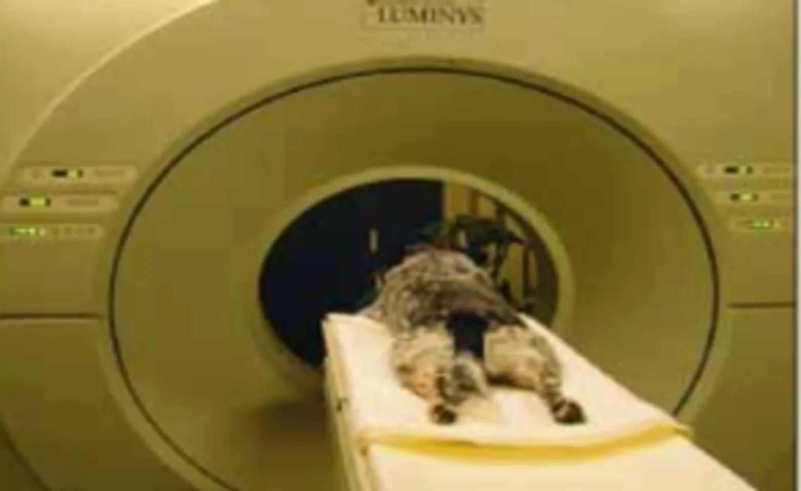
CT (computed tomography) is basically radiography performed in an axial plane (like bread slices). CT is excellent for evaluating bone detail and is ideal for visualizing bone tumors, spinal fractures, and discospondylitis. Soft tissues are better visualized than on plain radiographs, but the interpretation of subtle soft tissue lesions can still be difficult. In the case of type I disc extrusion in chondrodystrophic breeds, such as dachshunds, the extruded disc material is often mineralized. This mineralized disc material can be easily seen on a CT scan, and any lateralization of the disc material is also apparent. This is not necessarily true for larger dogs or dogs with type II disc disease, in which there is typically no mineralization of the disc. In these patients, a myelogram in combination with a CT scan is often needed.
Modern CT scanners are very fast, often completing a scan in a matter of minutes. A CT scan (without a myelogram) is non-invasive for the patient as well. Unfortunately, it is often impossible to know whether or not a myelogram will be needed prior to the scan.
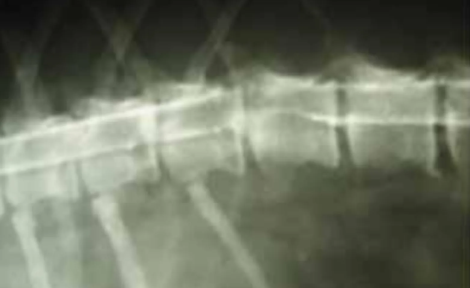
The photo at the top of this page shows disc material extruded into the right side of the spinal canal. A computed tomography (CT) image demonstrates extruded disc material on the right side of the spinal canal.
MRI (Magnetic Resonance Imaging) allows the best visualization of soft tissue lesions and is especially useful for diagnosing intramedullary spinal cord lesions, such as neoplasms, syringomyelia, arachnoid cysts, and even occasionally infarcts. It is also good at diagnosing disc disease in dogs. MRI is non-invasive but does require significant time to perform. It is the most expensive imaging modality, and because of the time required, scanning a large area (such as the thoracolumbar spine) in a large dog can be problematic.
